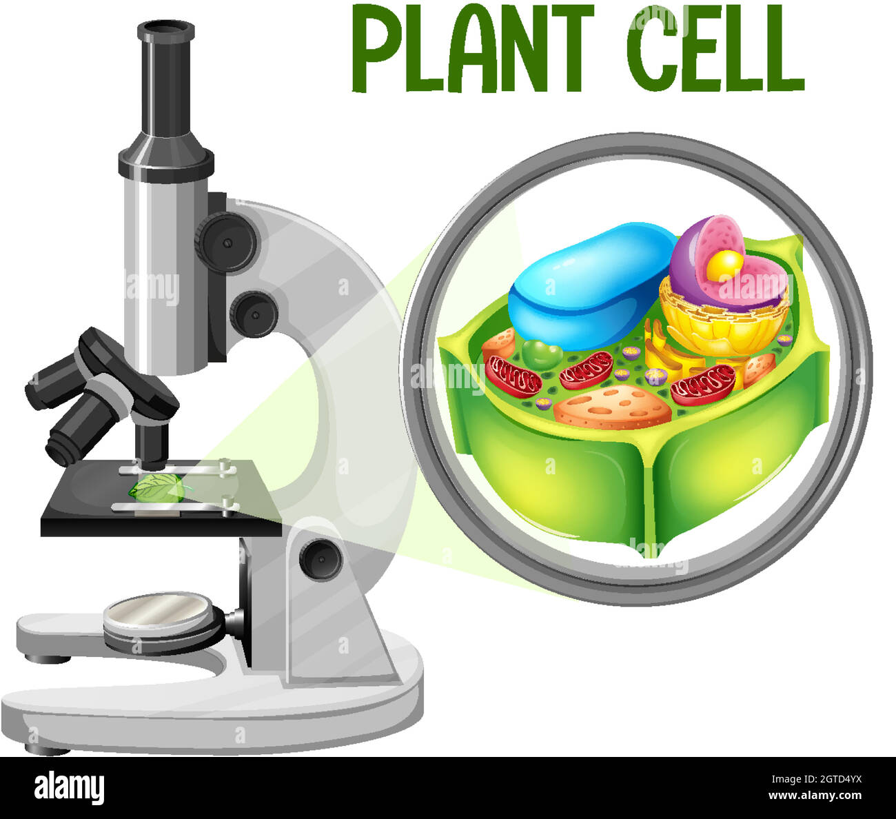
18+ Plant Cells Microscope
Web Microscopy-based imaging approaches allow researchers to analyze the dynamic localization of cellular components membrane remodeling events the morphology and function of organelles the structural features of proteins and molecular complexes and their interaction networks. Plants are multicellular photosynthetic eukaryotes whose cells produce cell walls composed of cellulose.

11 731 Plant Cell Microscope Images Stock Photos Vectors Shutterstock
Web Electron microscopes utilize focused electron beams rather than visible light and are capable of resolving or detecting much finer details than the light microscope.

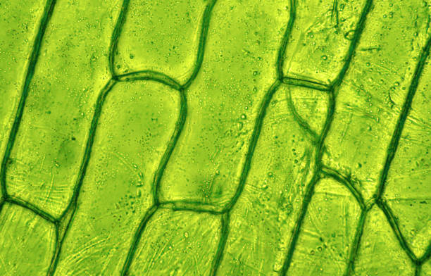
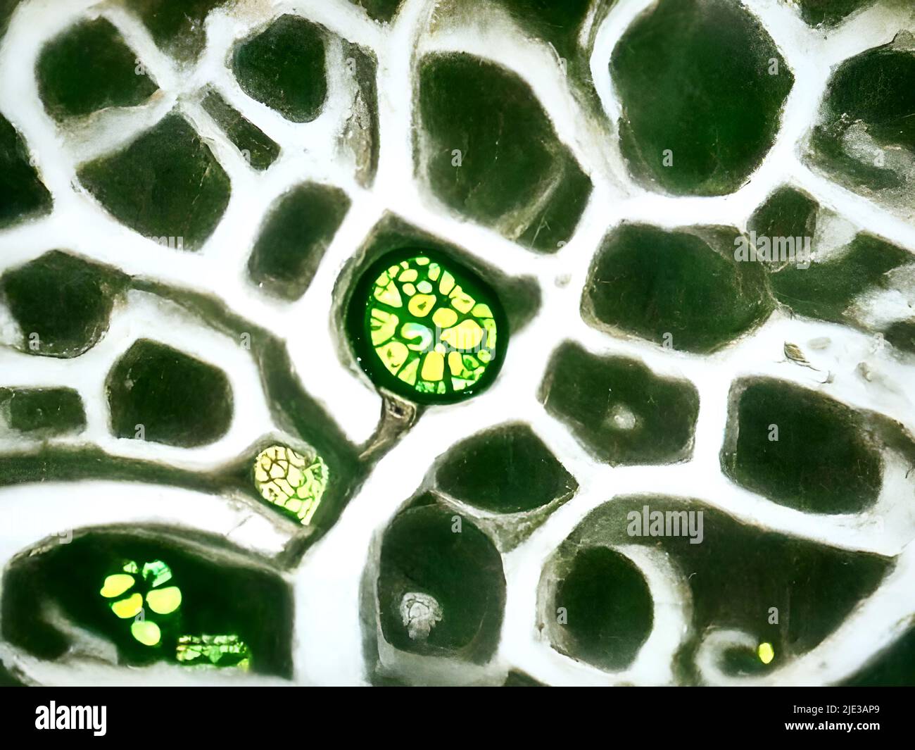
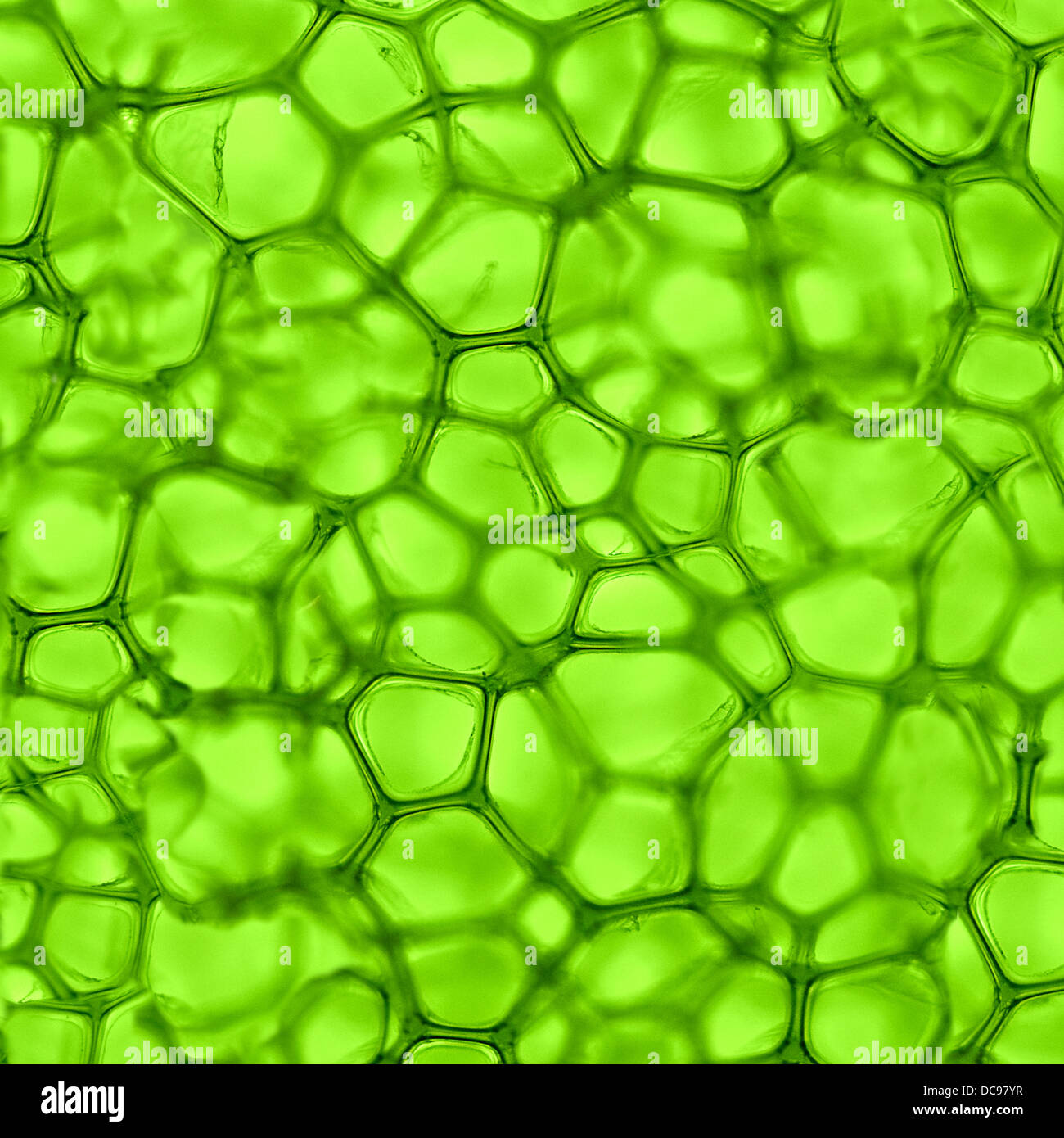
. In addition plant cells are characterized by vacuoles tonoplasts and plastids. Biologists typically use microscopes to view all types of cells including plant cells animal cells protozoa algae fungi and bacteria. Roots stems and leaves.
Web Gently scrape the inside of your cheek with a toothpick and swirl it in the dye on the slide. It is used primarily as a structural component in plant cell walls. Easy to handle fun and interactive the best plant cell microscope will open up so many different avenues of biology for you so lets get started.
Web Here we review some recent advances in live imaging of plant cells. In particular we discuss the solutions that plant biologists use to live image membrane-bound organelles cytoskeleton components hormones and. Web 1 LAB 1.
Living cells can be seen with phase-contrast differential-interference-contrast dark-field or bright-field microscopes. A new way to culture and image flowers is uncovering the processes that take place in reproductive cells buried deep in plants. In this activity students section plant material and prepare specimens to view under a brightfield microscope.
A diagram of a plant cell. LSFM includes microscopy methods with a unique geometry of imaging modalities which is conceptually distinct from classical widefield-based or pointillistic. Web The current efforts to study plant cells tissues organs and whole plants in developmentally oriented research by live-cell imaging are based on advanced microscopy platforms.
Cellulose is a carbohydrate composed of cross-linked chains of glucose molecules and it is difficult to break down. Microscopes Cells and Tissues Please bring your textbook to lab Objectives. Two types of electron.
Plants have three main organs. Draw 1-3 cells large enough to show the detail that you see in your lab manual. These microscopes are the compound microscope Figure 1 and the dissecting or stereo-microscope Figure 2Dissecting microscopes are commonly used for the observation of larger objects and generally have magnifications of less than 100x.
Plant cell culture is independent of. Web microscope Words. This lab will serve as an introduction to plant anatomy microscopy and the structure of a variety of plant tissues.
Web cell nucleus which is the control center of the cell and carries genetic information. Web Methods to enhance plant cell outlines vary in complexity from straightforward imaging of cell wall autofluorescence to lengthy multistep processing for three-dimensional analysis of tissue architecture by confocal laser scanning microscopy CLSM eg 1 3. Plant cells have a cell wall made of cellulose while animal cells have an enclosing cell membrane.
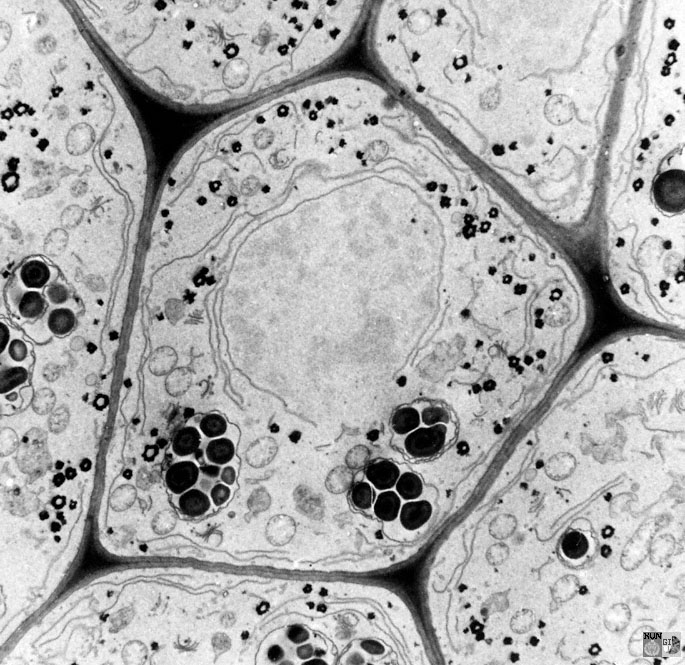
Label its cell membrane cytoplasm and nucleus. 18 June 2021 Biosensor imaging of a seedling measuring how the concentrations of the plant hormone gibberellin change as the plant grows. Web Scanning electron microscope SEM is one of the best analysis methods which has many uses in various fields including medicine biology biotechnology and industry.
Web In this review we discuss applications of cryo-ET to cell biology research on plant and algal systems and the special opportunities they offer for understanding the organization of eukaryotic organelles with unprecedently resolution. Web The limit of resolution of a standard brightfield light microscope also called the resolving power is 02 µm or 200 nm. Humans have been making use of plants for thousands of years.
Place a cover slip on the suspension and view at 1000X total magnification. Transmission and Scanning Electron Microscopy for Plant Protoplasts Cultured Cells and Tissues SpringerLink. Web September 30 2021 Scientists use the glowing properties of plant cells to capture stunning images by Botanical Society of America Formaldehyde fixation improves fluorescence patterns of.
Web Cells that have been fixed and stained can be studied in a conventional light microscope while antibodies coupled to fluorescent dyes can be used to locate specific molecules in cells in a fluorescence microscope. Plants cells differ from animal cells in that they have a cell wall which is glued to adjacent cells by the middle lamellae a large central vacuole and chloroplasts. Web Two types of light microscopes commonly are used in introductory plant pathology courses.
Looking below the surface in plants. Web New imaging methodologies with high contrast and molecular specificity allow researchers to analyze dynamic processes in plant cells at multiple scales from single protein and RNA molecules to organelles and cells to whole organs and tissues. Microscopy and stained specimens engage students visually as they learn about plant anatomy a topic covered in many biology and introductory science courses.
Both cell types contain cytoplasm. Web Plant cell microscopes are a must-have if youre looking to build on your collection or want a microscope for the fun of it. Spines hairs tendrils and thorns are usually modified.
Web We present a new large-scale three-fold annotated microscopy image dataset aiming to advance the plant cell biology research by exploring different cell microstructures including cell size. Carrie Metzinger Northover Bergmann Lab Stanford University. Was introduced as a well-known biotechnological method for producing SMs 16 17 proteins 18 and artificial seeds 19.
Web Confocal microscopy image of a young leaf of thale cress with one marker outlining the cells and other markers indicating young cells of the stomatal lineage cells that will ultimately give rise to stomata cellular valves used for gas exchange.

Biological Microscope Specimen Section Set Animal And Plant Cell Glass Slide Experiment Children And Students On Onbuy

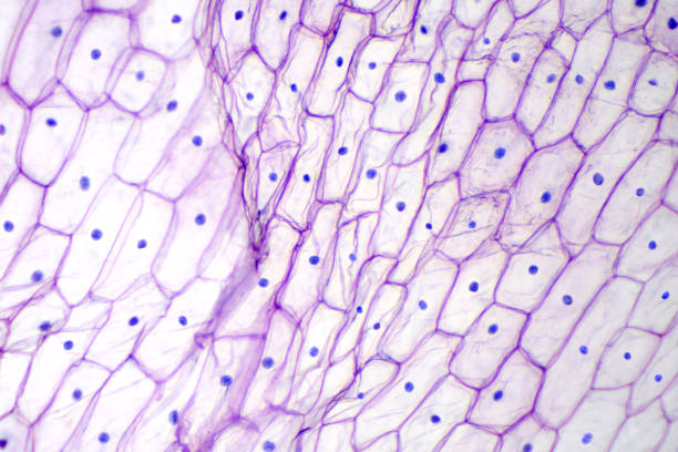
Microscope Image Of Plant Cells Stained For Nuclei Stock Photo Download Image Now Biological Cell Biology Biotechnology Istock

Green Plant Cells Under Microscope Seamless Vector Pattern Royalty Free Cliparts Vectors And Sto Plant Cell Microscopic Photography Things Under A Microscope
Plant Cell Microscope Hi Res Stock Photography And Images Alamy
6 700 Plant Cell Microscope Stock Photos Pictures Royalty Free Images Istock Plant Cell Wall
Plant Cells Microscope Hi Res Stock Photography And Images Alamy
Plant Cells Microscope Hi Res Stock Photography And Images Alamy
6 700 Plant Cell Microscope Stock Photos Pictures Royalty Free Images Istock Plant Cell Wall

Structure Of Animal Cell And Plant Cell Under Microscope Diagrams
6 700 Plant Cell Microscope Stock Photos Pictures Royalty Free Images Istock Plant Cell Wall

11 731 Plant Cell Microscope Images Stock Photos Vectors Shutterstock

Leapfrog Magic Adventures Microscope French Version Walmart Canada
6 700 Plant Cell Microscope Stock Photos Pictures Royalty Free Images Istock Plant Cell Wall

Cell Cross Section Under A Microscope Microscopic Cells Microscope Art Microscopic Photography

11 731 Plant Cell Microscope Images Stock Photos Vectors Shutterstock
Search In Gallery

Plant Cells Under Microscope Time Lapse High Res Stock Video Footage Getty Images